|
Free Pictures of Micrographs 1-40
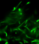
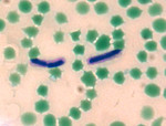
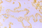
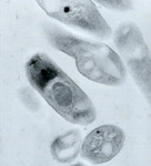
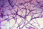
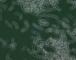
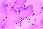
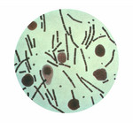
 Anthrax Direct Fluorescent Antibody (DFA) Cell Wall Stain
|
|
|
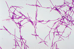
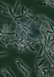
 Examining Fluorescent Micrographs
|
|
|
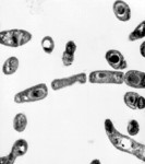
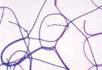
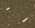 Bacillus Anthracis Indian Ink Capsule Stain
|
|
|
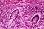
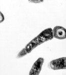
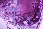
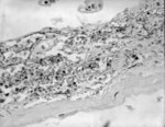 Bacillus Anthracis (Anthrax) in Meninges
|
|
|
 Bacillus Species Malachite Green Spore Stain
|
|
|
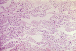
 Bacillus Anthracis in a Lung
|
|
|
 Strain of Bacillus Anthracis Bacteria
|
|
|
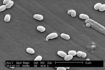
 Anthrax (Bacillus anthracis) Spores Micrograph
|
|
|
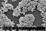
 Spores from Bacillus Anthracis (Anthrax) Bacteria
|
|
|
 Anthrax Transmission Electron Micrograph
|
|
|
 Bacillus anthracis (Anthrax)
|
|
|
 Transmission Electron Micrographic Image Of Bacillus Anthracis
|
|
|
 Anthrax Transmission Electron Micrograph
|
|
|
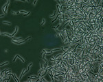
 Anthrax Bacteria Displayed During a Gram Stain Technique
|
|
|
 Bacillus Anthracis Gram Stain
|
|
|
 Positive Gram Stain with Bacillus Anthracis
|
|
|
 Bacillus Anthracis Spores
|
|
|
 Bacillus Anthracis Spores
|
|
|
 Bacillus Anthracis Spores Seen Under Phase Contrast Microscopy
|
|
|
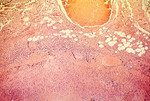
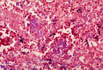
 Hemorrhagic Necrosis of a Lymph Node due to the Anthrax Disease
|
|
|
 Mediastinal Lymph Node from a Cynomolgus Monkey Infected with Anthrax.
|
|
|
 Necrosis Of Lymph Node Due To Anthrax
|
|
|
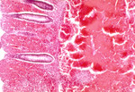 Histopathology Of Large Intestine In Fatal Human Anthrax
|
|
|
 Hemorrhagic Lymph Node Due To Inhalation Anthrax
|
|
|
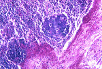
 Mild Meningitis with Hemorrhage due to Bacillus Anthracis
|
|
|
 Human Meningitis with the Presence of Bacillus Anthracis
|
|
|
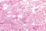
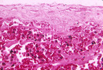
 Lung Tissue Infected with Bacillus Anthracis Bacteria
|
|
|
 Micrograph of the Fatal Inhalation of Anthrax in a Person
|
|
|
 Hemorrhagic Meningitis due to the Fatal Inhalation Anthrax
|
|
|
 Histopathology of Mediastinal Lymph Node in Fatal Human Anthrax
|
|
|
 Meningeal Hemorrhage due to the Anthrax Bacteria
|
|
|
 Anthrax Bacteria Taken From Heart Blood
|
|
|
 Bacillus Anthracis from Agar Culture
|
|
|
 Anthrax Taken from a Peritoneum Using a Hiss Capsule Stain
|
|
|
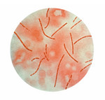
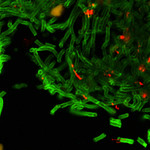 Green Anthrax Cell Walls and Red Anthrax Spores.
|
|
|
 Anthracis Direct Fluorescent Antibody (DFA) Capsule Stain
|
|
|
|
Next
|