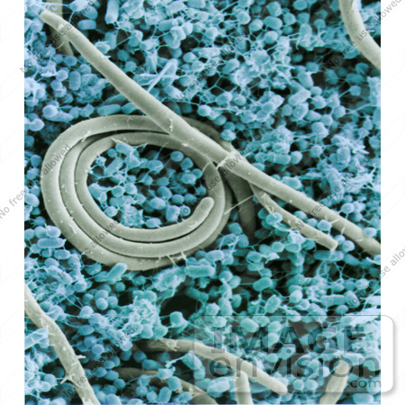
Stock Photography of a Micrograph of Salmonella Enteritidis Cells #26736


#26736 Stock Photography of a Micrograph of Salmonella Enteritidis Cells by JVPD
Royalty-free stock photo of a scanning electron micrograph of a mixture of small cells with filaments and very large cells that lack filaments. Salmonella enteritidis change shape as they grow. Small cells arise only during certain growth stages and efficiently contaminate eggs when the time is right.

Photo Credit: USDA/P.J. Guard-Petter/Stephen Ausmus [0003-0803-2418-1705] by 0003

Similar Images:

#14232 Picture of a Straw Itch Mite (Pyemotes tritici) by JVPD
#14228 Picture of a Two-Spotted Spider Mite (Tetranychus urticae) on a Rose Leaf by JVPD
#12960 Picture of a Red Palm Mite by JVPD
#6140 Stock Photography of a 1913 E. Leitz-Wetzlar Microscope by KAPD
#6142 Stock Photography of a 1913 Ernst Leitz-Wetzlar Light Microscope by KAPD
#6144 Picture of a 1913 E. Leitz-Wetzlar Microscope by KAPD
#6139 Stock Photography of a 1913 Ernst Leitz-Wetzlar Microscope by KAPD
#25802 Stock Photography of a Micrograph of Mites Infesting a Bee Trachea by JVPD
#14230 Picture of a Rust Mite (Aceria anthocoptes) by JVPD
#26737 Stock Photography of a Scanning Electron Micrograph of Fergusonina Fly Eggs and Associated Juvenile Fergusobia Nematodes in a Melaleuca Flower Bud by JVPD
#12967 Picture of a Red Palm Mite (Raoiella indica) by JVPD
#14227 Picture of the Yellow Mite (Lorryia formosa) by JVPD
#14229 Picture of a Yellow Mite (Lorryia formosa) by JVPD
#18653 Stock Photo of Possible Seeds of Planets, Green Crystals in Space by JVPD
More Microscopic Photos

Keywords

agriculture, bacteria, bacterium, blue, cell, cells, d036-1, electron micrograph, electron micrographs, filaments, microscopic, salmonella, salmonella enteritidis, scanning electron micrograph, scanning electron micrographs, usda